Ultrasound Physics & Knobology
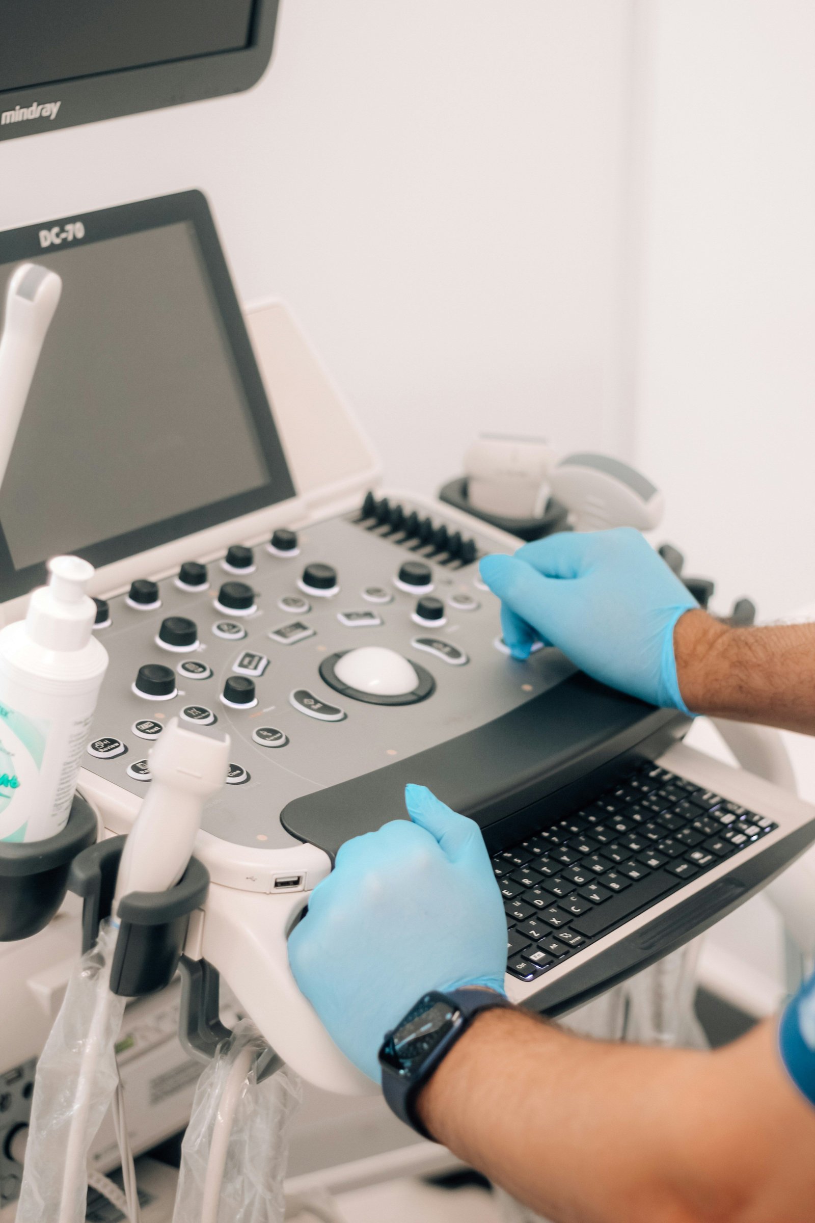
Configure 7.5–15 MHz linear probes for shallow tissue clarity. Set gain for anechoic vessels, center target depth, and align the focal zone.
See clearly. Cannulate with confidence.
Practical, supervised training on real-time high-frequency linear ultrasound for medical students, junior doctors, EM/ICU trainees, specialist nurses, and veterinarians.
Built for Human & Veterinary Clinical Practice
Every session prioritizes probe-in-hand repetition over slide decks. You leave with repeatable needle-tracking habits and a printed clinical reference manual.
Configure 7.5–15 MHz linear probes for shallow tissue clarity. Set gain for anechoic vessels, center target depth, and align the focal zone.
Differentiate compressible, thin-walled veins from pulsatile arteries and nerve bundles before skin puncture using compression and Doppler.
Apply the 1/3 lumen occupancy rule and 65% in-vein length rule (≥ 45 mm catheters) to reduce mechanical phlebitis and early dislodgement.
Drill the short-axis "walk down" technique at 30°–45°. Track the hyperechoic tip step-by-step until double-ring flash and full cannulation.
Prevent posterior wall penetration, recognize acoustic shadowing from tendons, protect median/radial nerves, and maintain sterile technique.
Complete a structured hands-on checklist covering vessel evaluation, dynamic tip tracking, and verified catheter flushing with instructor sign-off.
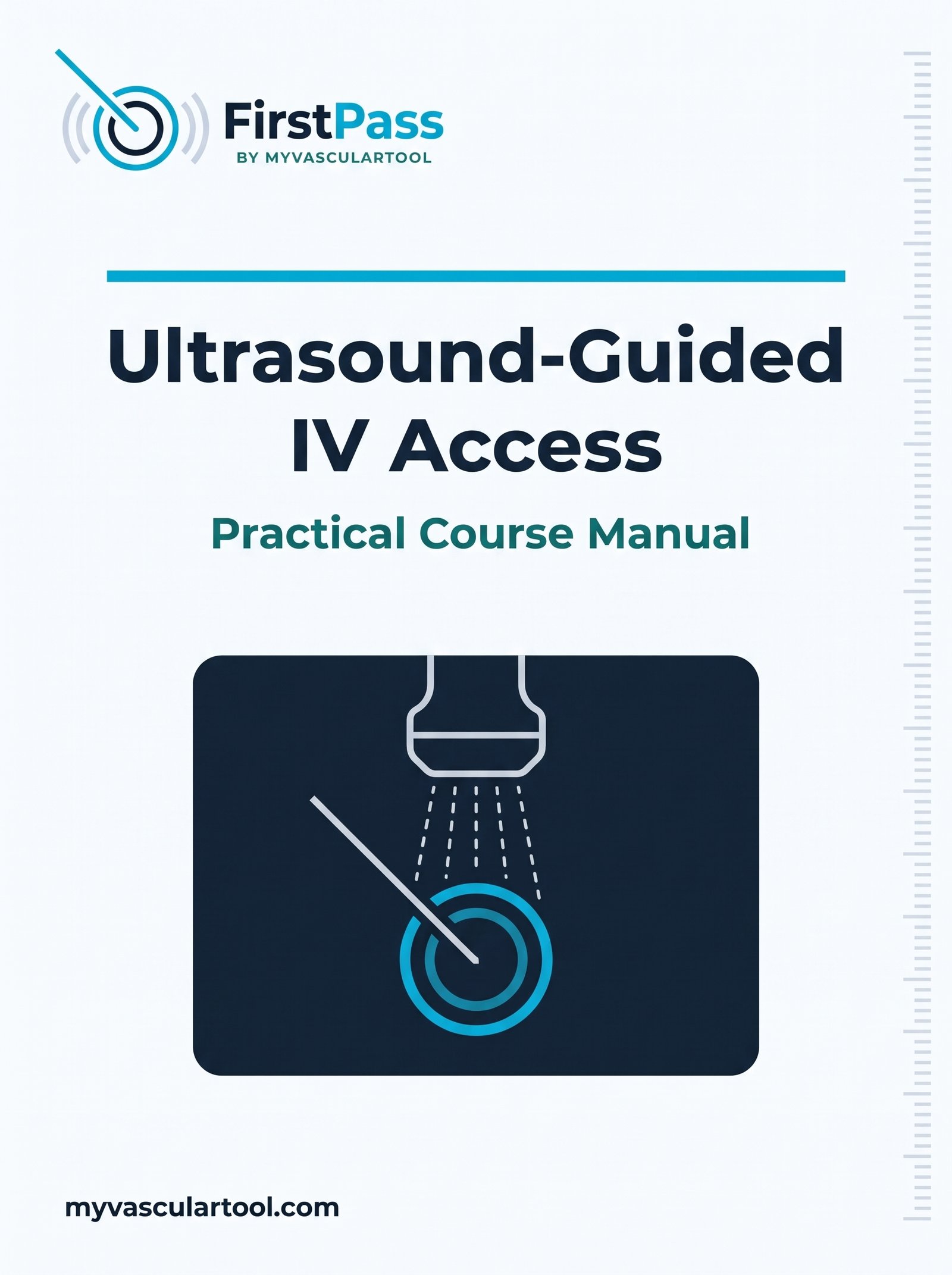
Take away our technical reference covering vein vs. artery sonography, catheter depth charts, and the 3-stage practical sign-off sheet for your training portfolio.
Reserve Your PlaceIndividual 1:1 sessions are hosted at our primary hubs in Norwich and Southend-on-Sea. For hospital wards, emergency departments, and veterinary practices along the A140 / A12 corridor—including Ipswich, Colchester, and Chelmsford—we offer on-site small-group training delivered directly to your clinical facility.
Transparent pricing for individual 1:1 sessions, with personalized quotes for small groups and clinical teams.
£150
Up to 3 hours • Dedicated Instructor
Focused 1:1 coaching tailored to your current skill level in Norwich or Southend.
Custom Quote
Tailored Duration • Norwich, Southend, or On-Site
Structured hands-on sessions for medical students, junior doctors, specialist nurses, or veterinary teams.
No. We start from probe orientation, gain, and depth setup before moving to short-axis needle tracking. Experienced clinicians can focus directly on deep or difficult vessels.
Yes. High-frequency linear ultrasound physics, vein-vs-artery differentiation, and dynamic tip tracking apply directly to veterinary vascular access.
In-person 1:1 and small-group sessions run in Norwich and Southend. On-site workshops for hospital departments and veterinary clinics can also be arranged.
No. All ultrasound systems, vascular access phantoms, needles, long catheters (≥ 45 mm), sterile gel, and printed manuals are provided.
Submit your preferred package, location, and availability. We respond within one business day to confirm your slot.
Primary Locations: Norwich & Southend (UK)
Direct Email: bookings@myvasculartool.com